More than Just For Pregnancies
Ultrasounds are most commonly associated with helping expectant mothers-to-be see their precious unborn babies. For most pregnant women, an ultrasound, also known as a sonogram, is the first glimpse of their child. For doctors and medical staff, ultrasounds play a critical role in monitoring the development of an unborn fetus and can help them visualize, identify, and diagnose many potential complications well before birth.
Ultrasound technology has much broader and greater applications than for just pregnancy. They can also be used to visualize, monitor, and image other vital parts of the body as well.
"Ultrasounds record images in real-time. By digitally recording a series of ultrasounds digitally, doctors and patients can better visualize changes over time within the body."
--- DR. CRISTIN DICKERSON, MD
What is an Ultrasound?
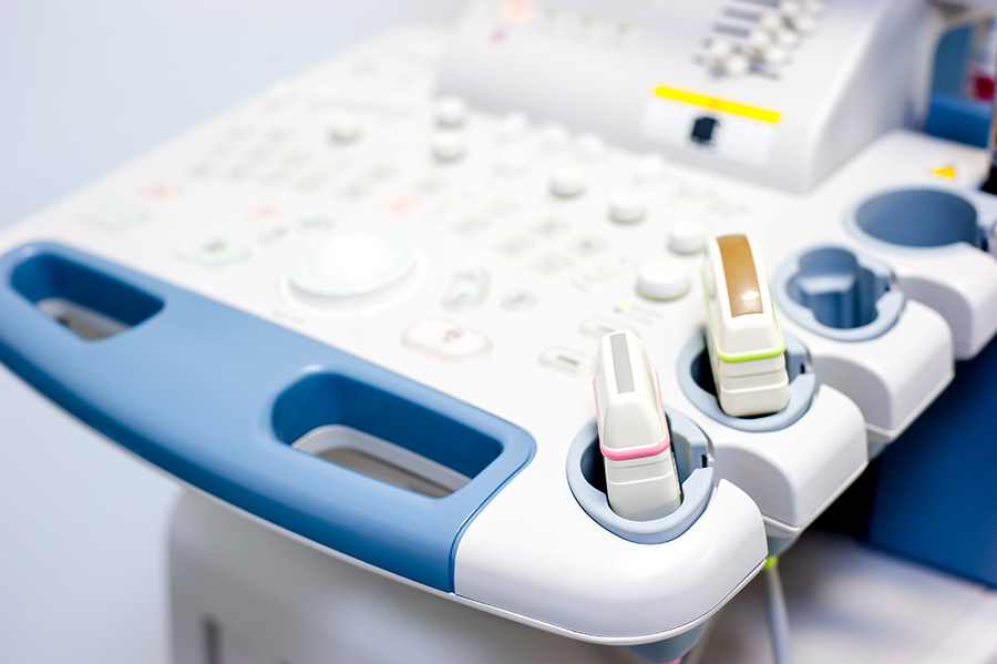
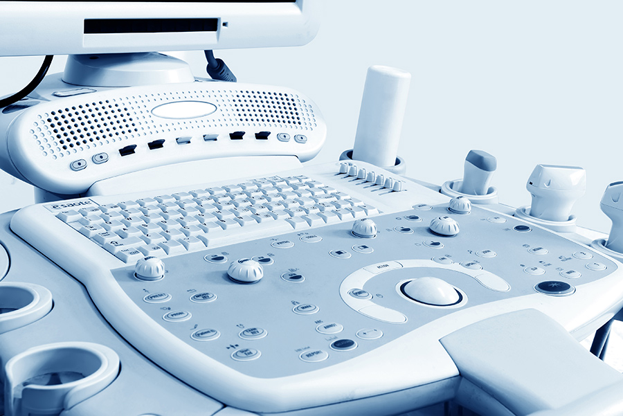
An ultrasound is a medical imaging procedure that utilizes sound waves to create an image of a person’s internal organs and anatomical structures. Ultrasound devices emit high-frequency sound waves, which are not discernible to the human ear, through a transducer which is set on a patient’s skin. The sound waves travel through the patient’s body and bounce off of internal organs and structures. Returning echoes are then translated into an on-screen image for the doctor and patient to see.
Ultrasounds record images in real-time. By digitally recording a series of ultrasounds digitally, doctors and patients can better visualize changes over time within the body.
What is the Ultrasound Procedure Process?
Each ultrasound procedure will progress differently and may require differing preliminary preparation depending on the organ or internal tissues to be examined. Some ultrasounds may need a general fast 8 hours prior while others may require significant liquid intake and a full bladder. Consult with the imaging clinic (i.e., Green Imaging) in charge before undergoing the procedure.
Typically, the first step in an ultrasound procedure is for the patient to remove their clothing, including jewelry and shoes, and change into a medical gown. Then, depending on the procedure, a patient may be asked to either lie down or sit in a private examination room. For surface-based ultrasounds and sonograms, a conductive gel may be applied at a location on the skin nearest the target area to help facilitate transmission of sound waves throughout the body. The technician or medical professional in charge then places a transducer on the skin. Sometimes, a wand may be used for specific applications. As echo waves from the transducer pass through the body, images are produced which are, in turn, sent to a radiologist for interpretation. The procedure is now complete, and the patient is free to change back into their regular clothing.
What Are The Different Types of Ultrasounds?
Doppler Ultrasound
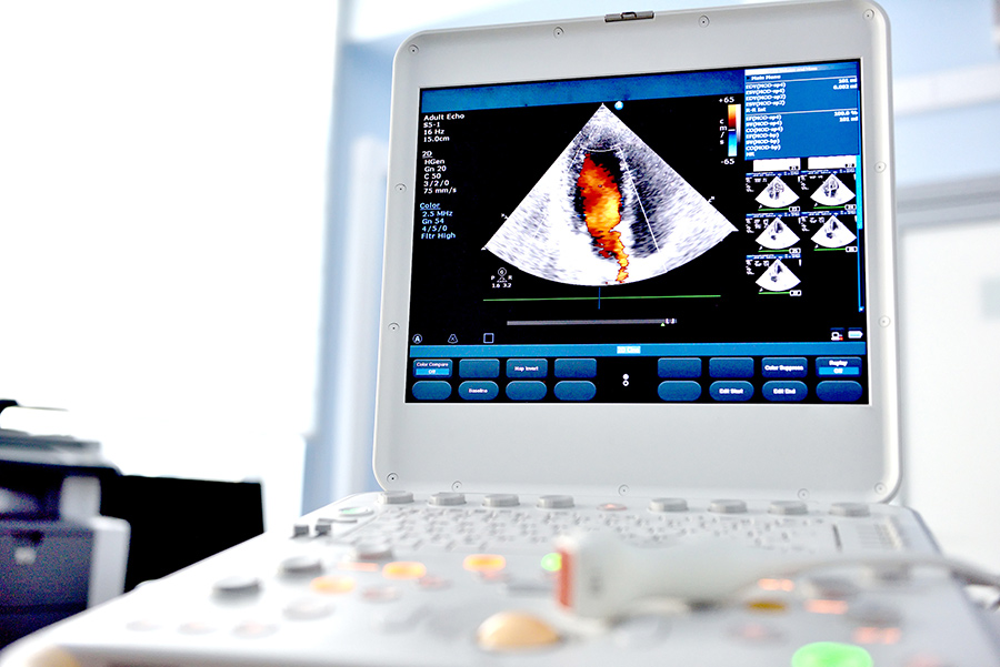
A Doppler ultrasound, often times simply referred to as a doppler, is a particular type of ultrasound used to evaluate blood flow in and around various organs and parts of the body. It produces valuable audio and visual data that can be easily graphed and quantified in other ways to help doctors evaluate and diagnose their patients. Dopplers are particularly useful for detecting the presence of dangerous blood clots, or the presence of deep vein thrombosis in susceptible patients and for diagnosing significant narrowing or dilatation of blood vessels.
Transrectal Ultrasound (TRUS)
A transrectal ultrasound (TRUS) is a diagnostic procedure utilizing an ultrasound probe to image the prostate gland and the surrounding tissue. The probe sends and receives sound waves and echoes through the rectal wall into the adjacent prostate gland.
Echocardiograms
Also known as an echocardiography, an echocardiogram is a diagnostic cardiac ultrasound used to visualize the heart. Echoes are used to create pictures and videos via a transducer, of your heart’s chambers, valves, walls and the blood vessels in motion.
Transesophageal Echocardiogram
Sometimes a standard echocardiogram cannot produce images of adequate fidelity of the upper chambers of a patient’s heart. In this situation, a transesophageal echocardiogram. What this means is that the echo transducer is inserted into a patient’s esophagus to get a clearer picture of the heart’s muscle and chambers, valves and outer lining (pericardium).
Bone Sonogram
As the name implies, a bone sonogram is an ultrasound designed to image a patient’s bone structures and is most useful in helping doctors identify signs of osteoporosis. Unlike a traditional x-ray, which is also very useful for visualizing bone structures, a bone sonogram does not produce radiation.
Transvaginal Ultrasound
A transvaginal ultrasound, also known as a female pelvic ultrasound, relies on a transducer wand which is inserted into the vagina to produce diagnostic images of the uterus, ovaries, and other reproductive structures.
3D Imaging
Unlike a traditional ultrasound which produces a 2D image, a 3D sonogram can create a live 3D representation of a baby within the mother’s womb. 3D ultrasounds can also make diagnosing specific birth defects such as cleft palate, easier.
4D Ultrasound
4D ultrasounds produce a series of 3D sonogram images over time to create a video.
Why You Might Need an Ultrasound
Generally, ultrasounds are used to image or visualize soft tissues within the body. These can be anything from a fetus and gestational sac to the large veins in a patient’s legs. Common reasons to undergo an ultrasound procedure include pregnancy and evaluations of other internal organs for damage or disease such as abdomen, pancreas, kidney, and heart. Another significant use of ultrasounds is for detecting blood clots and other disorders involving the arteries and veins.
Ultrasound Safety
Ultrasounds are non-invasive, generally painless, and incredibly safe. Unlike traditional x-rays and CT scans, ultrasound procedures do not produce or emit radiation. Nor do most require the injection of dyes or other contrast agents necessary for some MRIs. Ultrasounds are also widely available and usually less expensive than other imaging techniques.
The Green Imaging Difference
Choosing the right diagnostic exam for you takes a little time, patience, and research. Green Imaging can help. At Green Imaging, we are all about transparency and affordability. With Green Imaging you can save between 50 and 80% of your out-of-pocket costs for MRI, CT, ultrasounds, and other high-quality imaging services. Affordable MRIs start for as low as $250, compared with $1,600 at other imaging facilities in the Houston area.
Don’t pay secret rates for a diagnostic imaging exam. Go Green Imaging instead!


