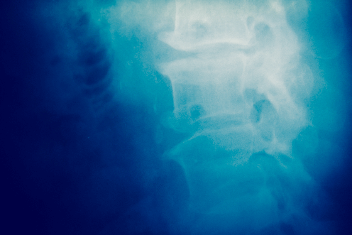
Spine Health
The human spine, also known as the vertebral column or backbone, is what holds our bodies up, gives us shape, supports our weight, and provides protection for the central spinal cord. When problems with the spine develop, such as disc herniations, bony spurs, spinal stenosis, and other medical issues, it is important for doctors to be able to see what’s happening beneath the skin, and muscles, behind organs, and inside of the spinal column itself. That is where myelograms come into play.
What is a Myelogram?
A Myelogram is a diagnostic imaging procedure specially designed to allow doctors to be able to see deep inside the spinal column. In order to do so, a myelogram procedure requires the careful injection of contrast dyes, or contrast agents, into the spinal canal. As you can imagine, this is no easy feat.
Myelograms are considered an outpatient procedure, which means they can be performed at a facility or clinic outside of a hospital.
What is a Myelogram For?
Because the spinal canal and the nerves therewith are hidden within the vertebrae of the spinal column, visualizing a patient’s spine with standard x-rays is not enough to produce the kind of high fidelity images and data doctors need in order to diagnose spinal deficiencies and diseases. Thus, a myelogram procedure is utilized to properly visualize the internal contents of the spinal canal.
This is accomplished with a combination of contrast dyes or agents, and x-rays or computed tomography (CT) scans carefully calibrated to allow visuals of areas where dyes are present. Myelograms don't replace MRI, they provide complementary information. They are also useful for determining the exact presence and degree of nerve root compression and better defining facet joint anatomy. Critically, myelograms allow spinal surgeons to accurately plan treatment and execute many surgical procedures.
What Does a Myelogram Procedure Entail?
Unlike a simple x-ray exam, a myelogram procedure requires a number of important steps to be executed safely and effectively.
1. Preparation
Patients are prepared prior to a myelogram procedure. Typically, this entails changing into a medical gown and laying on an exam table per the doctor’s instructions. Local anesthetics are applied in order to numb the tissues and prepare the patient to receive the necessary injection of contrast dyes and agents. Patients on certain blood thinning medications may also be asked to temporarily stop taking their medication prior to the procedure as a safety precaution.
2. Contrast Dye Injection
Once the patient is positioned laying on their back on the table, a needle is carefully guided into the spinal canal through the tough, protective dura without penetrating the nerves themselves. Dyes and contrast agents are then injected. These dyes mix with the spinal fluid and enhance images.
3. CT Scan Imaging
Once the dyes and contrast agents have been injected, the patient’s spine is then imaged with a computed tomography (CT) scan, or, in some cases, with multiple traditional x-rays from various angles. The dyes enhance the images produced by such exams allowing for greater detail, and thus, higher quality information for radiologists, surgeons, and other specialists.
4. Interpretation & Diagnosis
The images are then processed and sent to a trained and licensed radiologist who interprets the information. Comments and interpretations made by the radiologist are forwarded to the patient’s physician along with diagnoses and recommendations.
How Do I Prepare for a Myelogram?
Preparing for a myelogram is an important part of a smooth and successful procedure with no hiccups or side effects. Patients with pre-existing medical conditions, such as those taking blood thinners or those with diabetes, should always let their physician and the acting radiologist know about their conditions.
Patients should also inform their doctors about any allergies, particularly allergies to latex or chemicals found in contrast dyes. As one would imagine, failing to do so might lead to complications.
Additionally, it is recommended that patients abstain from eating or drinking (other than small amounts of water) at least 6 hours prior to the exam. Smokers should also avoid smoking as tobacco consumption has been shown to increase the odds and severity of certain side effects such as headaches.
Myelogram Concerns
The spinal canal is a highly regulated and sensitive part of the central nervous system. When spinal fluid pressure changes as a result of dural puncture, bad headaches can result. This is usually treatable with bed rest and over the counter painkillers.
Other potential concerns include accidental damage to or penetration of the spinal cord with the needle, possible infection of the spinal canal, and radiation concerns for pregnant women.
Pregnant women should never receive a myelogram procedure, as radiation from the imaging portion of the procedure can be harmful to the fetus.
Damage to the nerves themselves as well as infections is incredibly rare.
Overall, myelograms are an important and relatively safe diagnostic procedure that provide doctors and surgeons with valuable, visual information they need to make the best treatment decisions.


