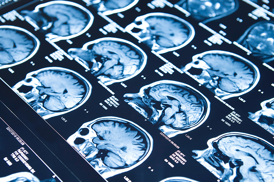
The Complete Guide To CT Scans
I am confident this is the most complete and comprehensive consumer guide you will find on the internet regarding CT scans.
In this guide, we will share with you our knowledge, life-changing stories, and experiences around medical imaging.
My name is Dr. Cristin Dickerson, founding partner of Green Imaging --- which is one of the leading full-service virtual medical imaging networks in the United States.
So if you're searching to learn as much as you can about CT scans, you will find value in our comprehensive guide.
DR. CRISTIN A. DICKERSON, MD
Contents
CHAPTER 1
What Is A CT Scan?
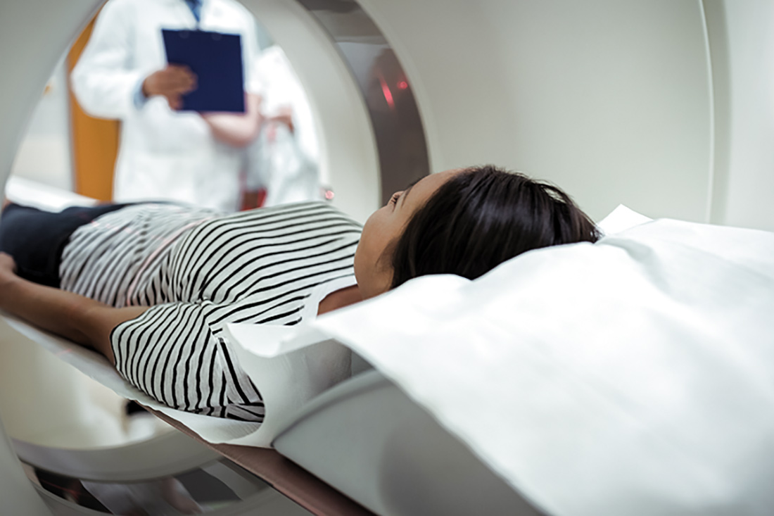
Computerized Tomography (CT) Scan Overview
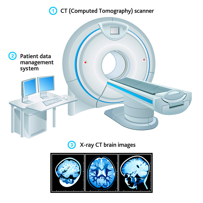
The computerized tomography (CT) scan procedure is now essentially a 3D version of the traditional x-ray examination. In essence, a CT scan is an x-ray volumetric acquisition of data from a part of the body. The images are reformatted by computers (hence computerized) to create standard cross-sectional and 3D images.
Brief Medical History of the CT Scan
Computerized tomography (CT) scan technology has been well established for over half a century. The first commercially available CT scan equipment was conceived in 1967 and installed in 1971, although the basis for the technology was demonstrated on several occasions well before that time. The roots of CT scan technology go all the way back to 1917 with the development of radon transform mathematics.
The first CT scan machines were primitive by modern standards. The first machines could only take cross-sections of a patient’s brain. Patients had to stick their heads into a water-filled tank which was necessary to lower the dynamic range of the x-ray radiation for detection purposes. Images produced by this early CT scanning machine could only produce images with a resolution of about 80 by 80 pixels. For comparison, the average resolution of a computer screen today is 1366×768. Modern CT scanning machines can resolve images 2048 by 2048 pixels.
Not long after the introduction of the first CT machines, researchers discovered how to make working scanners without the need for a tank of water. Over time, CT machines have rapidly advanced in terms of operating speed, slice count, image quality, and patient comfort. However, the fundamental idea behind CT scans has remained the same.
Types of CT Scans
CT scans can be employed for nearly any part of the human body. Depending on which part of a patient’s body is targeted for imaging, the exact procedures and techniques may differ somewhat. However, the overall concept remains the same.
Creating three-dimensional and cross-sectional images of the body part in question can help doctors better visualize hidden conditions within a patient's body, leading to more accurate diagnoses and better outcomes as a result.
The most common types of CT scans include CT scanning of the head, CT scans of the neck, CT scans for coronary calcium, CT angiographies (CTAs), CT scans of the chest, CT scans of the abdomen and pelvis, and CT scans of the spine.
CT Scan of Head
CT scans are well suited for imaging the head and brain. The very first commercial CT scanning machine could only image a patient’s head. Modern head scans can provide doctors a bevy of high resolution and accurate information on a patients head, including the soft tissues of the brain, the hard tissues of the skull, as well as the many intricate blood vessels in and around the head. The reliability of a CT head scan has made the procedure one of the standard diagnostic tools in hospitals worldwide.
A CT scan of the head can help doctors locate fractures in the skull bone, contextualize brain injuries, and detect bleeding or blood clots in the head. Head scans can also be used to visualize the extent and scope of traumatic damage to the face and head and help guide maxillofacial surgeons during reconstructive surgery. CT scans are also used to detect brain tumors, assess brain ventricles, assess disease in the paranasal sinuses, evaluate skull malformations, and even assess aneurysms through a CT angiography.
CT Scan of Neck
CT scans of the neck evaluate the nasal and oral cavities for disease, evaluate the size and lesions of the salivary and thyroid glands, evaluate the airway, and evaluate the lymph nodes in the neck.
CT Scan for Coronary Calcium
These scans are simple non-contrast scans of the heart, acquired to assess for the presence of atherosclerotic calcification in the coronary arteries. Computer software is used to quantify that volume of calcium. The software then compares that volume to a database that allows determination of a patient's risk of future adverse cardiovascular events like heart attack and stroke. It is in fact, the best noninvasive test we have to predict that risk and usually only costs a few hundred dollars.
CT Angiography (CTA)
CT angiographies (CTAs) are designed to visualize blood flow in regions of the body. This is extremely useful for a litany of important medical reasons. CTAs can be used to visualize and examine pulmonary embolisms, assess brain aneurysms and arteriovenous malformations, display highly accurate information on the renal arteries, and detect atherosclerotic disease. The application of CT technology for angiographical purposes has revolutionized how modern doctors visualize, detect, and treat all sorts of diseases from heart disease to stroke.
CT Scan of Chest
CT chest scans are often recommended after a traditional chest x-ray has detected something that may require further investigation. The detailed cross-section produced by a CT chest scan can either confirm previous findings and provide additional detailed information, or, determine that the earlier results were false-positive (artifactual).
CT chest scans are also particularly useful for the monitoring of individuals who are at a higher risk of developing lung cancer. Low radiation dose techniques have been developed for these scans to minimize the radiation risk of having annual CT scans.
CT Scan of Abdomen and Pelvis
Abdominal and pelvic scans using CT technology is an excellent diagnostic imaging technique for identifying a variety of issues including identifying abscesses in the abdomen or pelvis, inflamed bowel, diverticulitis, appendicitis, and assessing the presence and extent of cancers. CT scans of the abdomen are particularly good at imaging and visualizing liver, spleen, pancreas, adrenal glands, kidneys, and gastrointestinal (GI) tract.
CT Scan of Spine
CT scans are often recommended for diagnostic imaging of the spinal column due to the complexity of the human spinal column, including the presence of a wide variety of tissue types that include bone tissue, nerve tissue, muscles, blood vessels, and soft tissues, in a relatively condensed region of the body.
The intricate shapes and details of internal structures, such as delicate vertebrae bones, can be visualized in high resolution with CT technology. As a result, CT spine scans are commonly used to detect spinal injuries, tumors in the spinal column, metastases of other cancers, vertebral fractures, infections, and the presence of degenerative diseases such as arthritis.
CHAPTER 2
CT Scan: Uses, Side Effects, Process, & Results
A CT scan is generally used as a clarifying diagnostic tool after a patient’s doctor has reason to suspect the presence of disease and a standard x-ray diagnostic has proven inconclusive and occasionally as a screening exam for lung cancer and coronary artery plaque.
In some cases, such as a head or neck injury, a CT scan may be the first diagnostic ordered to identify and diagnose bleeding or swelling in the brain quickly.
Examples of when a CT scan may be in order include:
Inconclusive x-rays
Head injury
Spinal injury
Small or fine fractures
Imaging of complex structures
Imaging of organs and other soft tissues
Imaging for surgical planning purposes
CT Scan Side Effects
CT scan technology is built on x-ray technology. To produce detailed three-dimensional cross-sectional data for imaging and diagnostic purposes, a CT scan is like x-rays obtained at many different angles. As we know, x-rays produce strong gamma radiation that can damage cellular DNA and at high doses may cause cancer.
CT scans pose the same small but not insignificant radiation danger for patients. The risks of developing cancer as a result of a CT scan are about 1 in every 2,000 CT scan patients. While this number is small, repeated scans can increase a person’s risk of developing cancer.
The effects of radiation exposure can build up gradually over a long period. That’s why doctors only prescribe a CT scan only when necessary. To reduce a patient’s radiation exposure, CT scans are typically reserved as a clarifying diagnostic after a standard x-ray has detected something out of the ordinary.
As a result of the potential dangers of ionizing radiation, children and pregnant women are usually prohibited from receiving CT scans unless the potential benefits outweigh the long term danger posed by radiation exposure.
While radiation is a real concern, it is essential to understand that the risks are still minimal. Proper protection and operation are essential. Patients concerned about the potential side effects of CT scans should consult with their doctor.
3 Steps of a CT Scan Process
Preparation for a CT Scan is very straightforward with a few exceptions. In general, the only preparation needed for a CT scanning procedure is to change into a medical or hospital gown. Medical gowns promote general cleanliness and hygiene and ensure that no unintended items, such as keys or tiny fibers of clothing, make it to the CT scanning table.
Some CT scan procedures, such as an angiogram, may require the injection of scan-specific contrast agents and imaging dyes. In the case of an angiogram, patients must receive an injection of iodine which helps the CT scan visualize essential parts of the heart and blood vessels. Some CT scan procedures may also require patients to refrain from eating or drinking during a specific period leading up to a CT scan procedure. These important preparatory steps ensure the accuracy and efficacy of the CT scan.
Other scans require a patient to drink an oral contrast agent over a period of time prior to the exam to opacify the bowel loops for their better evaluation.
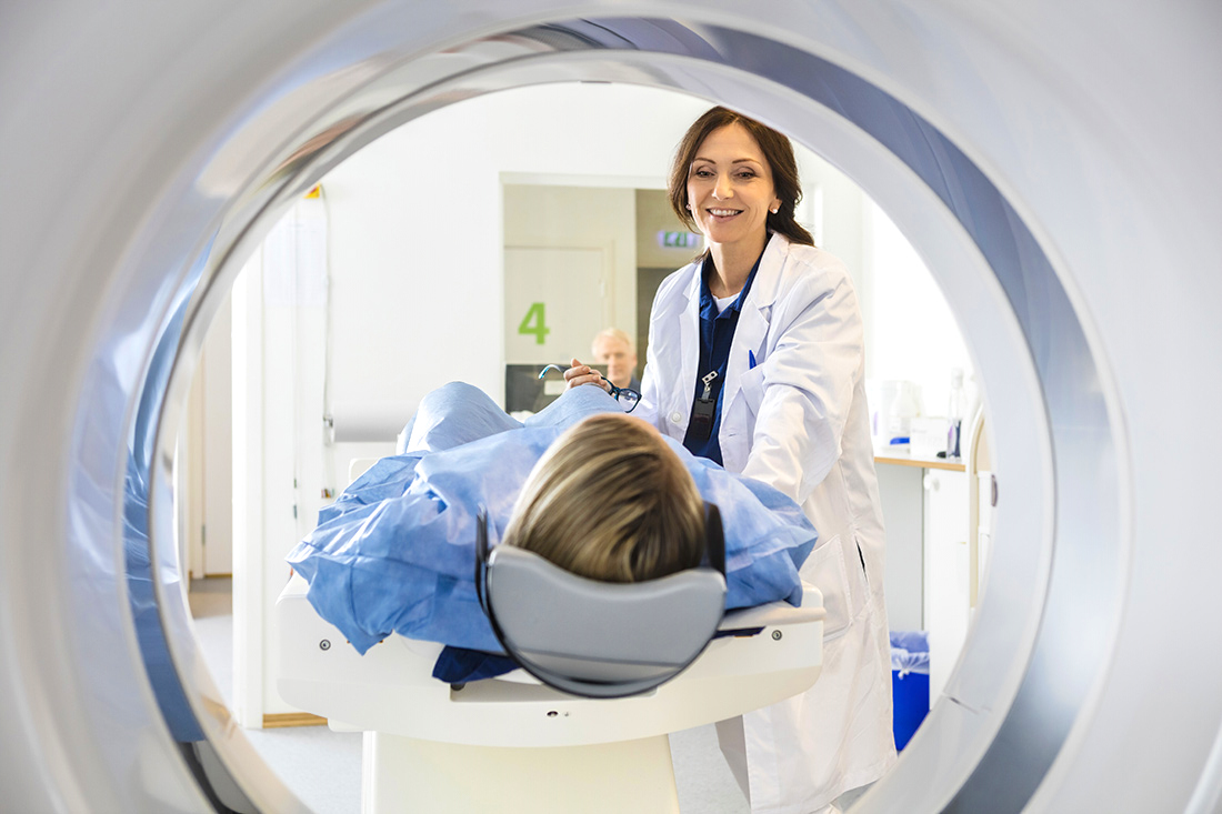
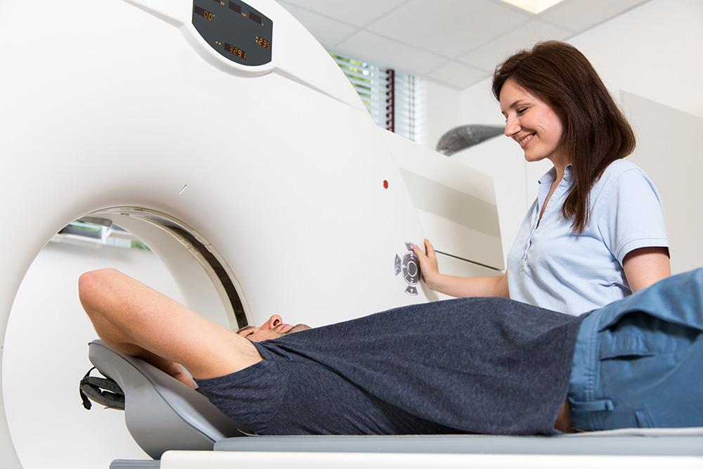
During a CT scan procedure, patients will be asked to lie down on the scanning table in a position most conducive for the particular CT scan required. Sometimes patients will lie on their back while other times they may be necessary to lie on their side.
Once in place, the technologist or radiologist will leave the room and enter an adjacent control room. The table slides beneath or into the CT scanning equipment.
Once in place, the CT machine begins taking cross-sectional images of the patient’s body. The table on which the patient lies may move slightly to take different images.
It is vital that a patient remains still during the CT scanning machine's operation.
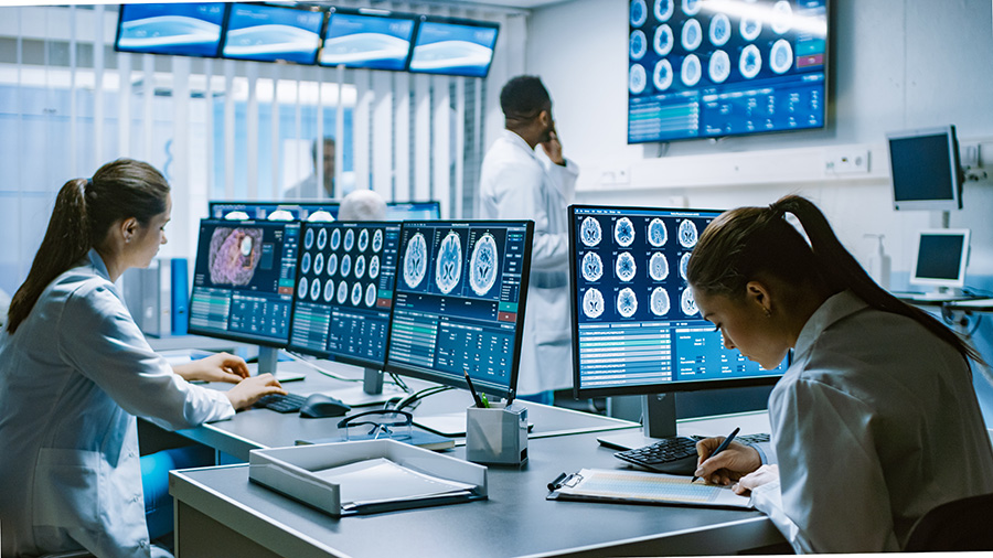
After the procedure patients change back into their street clothes and can go about their day, the images from the CT scan are typically presented to a radiologist who then interprets the data. Based on the data provided, the radiologist will then provide a patient's physician with a report describing the findings along with professional recommendations and diagnoses.
CT Scan Results
Radiology reports, as well as the actual computed tomography images themselves, can be difficult for a non-professional to read or understand. Typically, the report, images, and professional recommendations or diagnoses are not sent to a patient, but rather to the patient’s primary care physician or another specialist who will then provide the information to the patient and correlate it with lab and clinical findings.
Reports are often chock full of medical terminology and healthcare jargon and are primarily intended for other medical professionals.
Generally, radiology reports are broken down into six sections:
1. Type of Exam
Here you will find the type of examination that was provided, such as a CT scan, MRI, or another diagnostic examination.
2. Clinical Information
This is where a patient’s clinical symptoms are communicated to the radiologist. Having a concise but thorough patient history here also helps the radiologist to interpret the data more accurately.
3. Comparison
This section denotes other examinations and previously obtained data that may inform the radiologist’s interpretation.
4. Technique
This is where a radiologist describes the exact methodology used for the examination and other technical details.
5. Findings
This is by far the lengthiest part of any radiology report. Here, the radiologist will go into detail about his or her observations including whether or not specific parts of the body that were analyzed are normal or abnormal.
6. Impression
In this final section is where all the data and proceeding information is synthesized to generate a coherent diagnosis or diagnoses. Many important interpretations and answers to the clinical question(s) will be found in this section of a radiology report.
CHAPTER 3
CT Scan vs. MRI
Computerized Tomography (CT) Scan
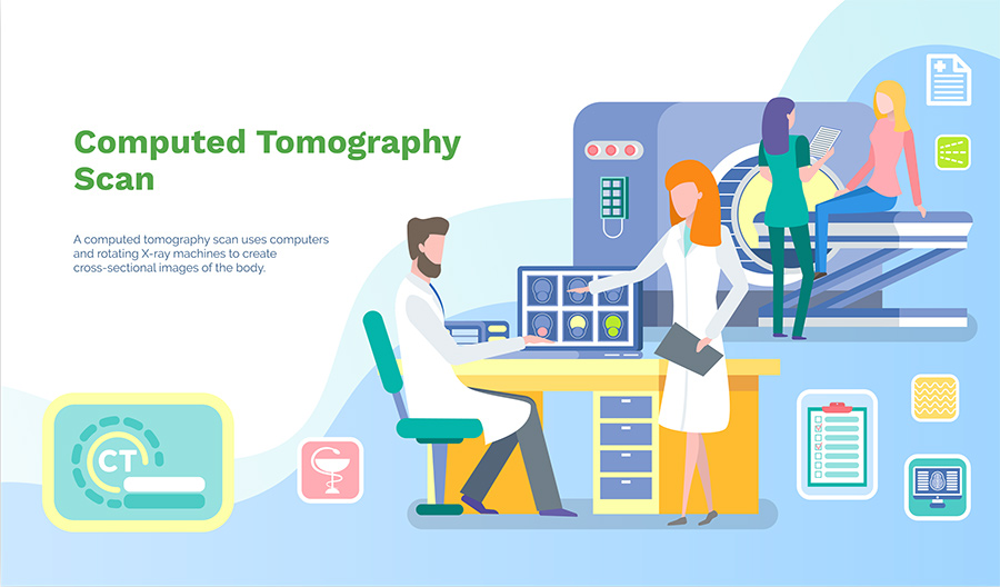
The computed tomography (CT) scan is one of the most commonly prescribed diagnostic imaging procedures in the world. In the United States, over 70 million CT scans are done every single year. Nonetheless, this common diagnostic examination often gets confused with another widespread diagnostic test: the magnetic resonance imaging (MRI) examination.
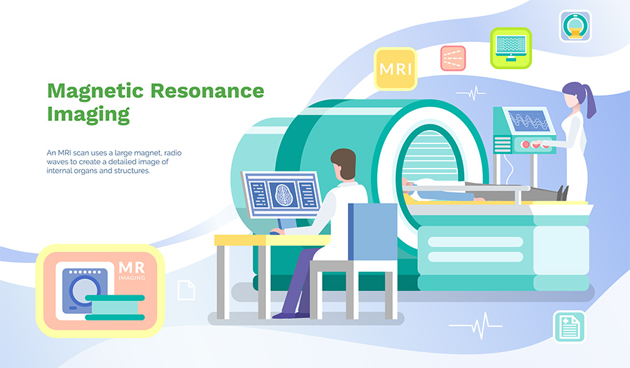
In the United States alone, over 34 million MRI exams are carried out every year. In total, these two exams account for a majority of the advanced diagnostic imaging examinations executed in any given year.
While both CT scans and MRI exams serve a similar important diagnostic function, the two imaging procedures couldn’t be more different. Not only do the underlying mechanisms and machinery differ between the two diagnostic tests, but the ways in which they are used diverge as well.
To understand the vital role CT scans play in modern health and medicine, it is essential to know how CT scans compare with other important diagnostic imaging tools such as MRI exams.
CT Scan & MRI Exam Similarities
Both CT scans and MRIs are diagnostic imaging techniques that are designed to help doctors identify, locate, and clarify the presence of existing diseases or injuries. Both procedures accomplish this through the generation of imaging data which is then interpreted by a radiologist.
The preparatory steps patients must undergo before a CT scan, or MRI exam is nearly identical. In both cases, patients must change into a lab-certified medical gown and remove all jewelry and accessories. While the exact reasons for doing this is different for each examination, the goal in both cases is to ensure patient comfort and safety.
Both CT scans and MRI’s may involve the injection of contrast dyes or radiocontrast agents to aid in creating highly specific and detailed images for particular purposes. Radiocontrast agents are used in CT scan applications for routine studies and more complex procedures such as angiography, enterography, colonography, and arthrography.
CT Scan & MRI Exam Differences
One of the most significant differences between a CT scan and an MRI is the use and application of radiation. CT scanning technology utilizes a series of powerful x-rays to generate three-dimensional data.
X-ray imaging produces radiation that can be harmful to patients. Although patient exposure to harmful x-ray radiation is relatively minor, it is not insignificant. Nonetheless, doctors do not recommend CT scans to pregnant women or children except in cases where the potential risks of radiation exposure are outweighed by the potential benefits provided by a CT scan.
MRI machines, on the other hand, do not emit any radiation whatsoever. However, MRI’s are known to emit loud, uncomfortable noises and may not be an option for some patients with pacemakers and defibrillators, or patients with implanted devices containing ferrous metals. CT scans, on the other hand, are safe for all medical devices.
The means and methods by which CT scans and MRI tests generate imaging data are entirely different. CT scanners emit a series of narrow X-ray beams through the human that move through an arc. The data acquired is used to produce a detailed three-dimensional model.
MRI machines do not use radio imaging to generate imaging data. Instead, MRI’s utilize the tiny magnetic fields generated by each atom in our bodies. The MRI machine creates a powerful magnetic field which forces the protons in a patient's body to align with it. The MRI technician then introduces a radio-frequency pulse that disrupts this magnetic alignment. Once the pulse ends, the protons in a patient’s body realign releasing detectable electromagnetic energy. Depending on the type of tissue, such as muscle tissues, fat, or organ tissues, differing amounts of electromagnetic energy are released. A computer parses this information and generates a digital image of the patient’s body based on the electromagnetic energy signatures detected.
In general, CT scanning procedures are executed must faster than MRI exams. A typical CT scan will take no more than 10 to 20 minutes to perform.
In comparison, the average MRI exam can take from 30 minutes to as long as a couple of hours. That is because many MRI exams are typically executed in 5-minute blocks between which the machine must be reset and recalibrate for the next block.
Furthermore, the data gathered by MRI machines are usually much more complex and detailed. When this relative complexity is combined with the need to re-image parts of the body two or three times with different MRI settings to capture information on different tissue structures, MRI procedure times can balloon quickly.
CT scans and MRIs are used to image very different parts of the body. Where one technique may be lacking in regards to resolving important imaging data, the other may excel and vice versa.
For example, CT scans are extremely good at visualizing the head and facial structures as well as the bones in the body. They are very useful for finding fractures in bone that would be otherwise invisible to a standard x-ray examination. CT scans also have the advantage of being very fast which make them invaluable for imaging patients with traumatic brain injuries who need treatment very fast.
MRIs are much better than CT scans at visualizing and resolving soft tissue details such as organs and complex structures such as spinal vertebral columns. The fine degree of soft tissue contrast offered by MRI allows doctors to see fine detail like individual tendons and nerves. MRIs are also extremely useful for identifying tumors in a patient’s body.
Ultimately, no single imaging procedure will work for every patient’s needs. Some patients may require both CT scans and MRI exams to provide doctors with the most information to make a diagnosis or to treat a disease. What matters is that the medical questions being asked can be answered. Your doctor and radiologist can help decide which procedure will work best for you depending on the clinical questions that need to be answered.
CHAPTER 4
CT Scan With Contrast vs. CT Scan Without Contrast
Computed Tomography (CT) Scan
A Computed Tomography (CT) scan is a common diagnostic imaging procedure. Millions of patients undergo this essential imaging examination every year to help doctors see into the inner working of their patients’ bodies.
However, while incredibly useful, CT scans are not a perfect imaging tool and have certain limitations. CT scans may, for example, have difficulty resolving extremely fine details in complex soft tissues and organs. Abdominal CT scans and angiograms, for example, can be notoriously difficult to read without the aid of substances and imaging aids collectively known as contrast mediums, contrast agents, or contrast dyes.
What is a CT Scan Contrast Dye?
Contrast dyes, also known as contrast mediums or contrast agents, are substances designed to improve the imaging capability of a standard CT scan by increasing the contrast of structures or fluids within the body. What this means is that these dyes or contrast agents are expressly introduced into the body to make certain parts of the body stand out in CT scan images.
Contrasts are consumed or injected where they strategically collect in specific parts of the body depending on the formulation. One dye may be designed to increase the contrast of abdominal tissues. Another dye may be designed to help the cardiovascular system stand out more with surrounding tissues. In both cases, the goal of the contrast agent is to highlight the specific structures, organs, tissues, and fluids that the doctor wants to examine.
The contrast dyes themselves can be made from a variety of materials, including iodine and barium. The exact dye used for each CT scan procedure that calls for contrast materials will depend on the precise organ or internal structure that needs to be imaged.
Types of Contrast Materials
The vast majority of contrast agents are either iodine or barium-based. Contrast dyes can also be subdivided by their method of delivery: oral contrast, rectal contrast, IV contrast, or intrathecal contrast.
Typically iodine or barium-based, oral contrast dyes are most commonly used when imaging a patient’s bowels. Oral contrast is also widely used in abdominal and pelvic CT scans. The patient drinks this contrast and it is not absorbed by the bowel like food and water are. It stays in the bowel and enables a radiologist to differentiate between the bowel and other organs and to better assess the bowel for abnormalities.
Rectal contrast, or delivery of contrast materials through the rectum, can be used to deliver contrast to a patient’s lower bowels. This is typically done when lower bowel pathology is suspected, especially a leak in the lower bowel.
Intravenously introduced contrast dyes are used to aid in visualizing and imaging vascular structures within the body.
Contrast dyes can also be directly injected into the spinal canal (intrathecal) to evaluate for spinal diseases and identify cerebrospinal fluid leaks.
For What Are Contrast Agents Used?
Contrast dyes are used to drastically increase the contrast of specific tissues or internal structures in a CT scan image thereby making them more apparent and more visible.
Examples of specific CT scanning procedures that utilize contrast dyes include:
Brain CT and circle of Willis CT angiography
Neck CT and carotid CT angiography
Chest scans and pumonary embolus and aortic aneurysm CT angiography
Myelography
Abdominal and pelvic CT and CT angiography
Runoff lower extremity CT angiography for vascular disease
How Do Contrast Materials Work?
Contrast agents contain either iodine or barium which x-rays cannot pass through. The use of contrast materials to produce higher contrast images is known as opacification.
Contrast dyes, whether they are administered orally, intravenously, rectally, or intrathecally, isolate to specific tissues making those tissues opaque, and therefore more visible to the CT scanning machine.
Contrast Precautions
For the vast majority of patients, CT scanning procedures that utilize contrast agents are perfectly safe. However, it is essential to take precautions when considering a CT scan. Some people may have dangerous allergies to common contrast materials such as iodine. Others may have complicating factors such as systemic diseases like heart disease or diabetes.
In particular, women should notify their doctor or radiologist if they are or might be pregnant. CT scans themselves, as well as certain contrast dyes, can be harmful to fetuses. Likewise, mothers of young babies should avoid breastfeeding their children at least 24 to 48 hours after a CT scanning procedure to avoid exposing their children to contrast agents.
Certain at-risk groups include those with:
Cardiovascular heart disease
Asthma
Allergies
Dehydration
Previous kidney failure
Renal disease
Contrast materials from an earlier session in their bodies
Any prior allergic reaction to iodinated contrast
CT Scan With Contrast vs. CT Scan Without Contrast
CT scans that require administration of contrast dyes often also need more preparatory work from both patients and doctors than a standard CT scan that does not involve the use of contrast mediums.
First and foremost, doctors must evaluate a patient to ensure that contrast dyes will be safe for them. This usually entails renal function laboratory testing. It is essential for patients to communicate with their doctor about any allergies, such as a known allergy to contrast materials such as iodine. Doctors will also need to evaluate if certain systemic factors, such as heart disease, diabetes mellitus, or age might make the administration of contrast agents unadvisable.
If contrast dyes are called for, specific preparatory steps must be taken. Some CT scanning procedures may require that patients refrain from eating or drinking for a certain amount of time before an exam. Other contrast dyes are very time sensitive and require precise timelines.
If your CT scanning procedure calls for the use of contrast materials, be sure that you read and understand your doctor’s instructions very carefully.
CHAPTER 5
What Does A CT Scan Show?
Computed Tomography (CT) Scanning Technology
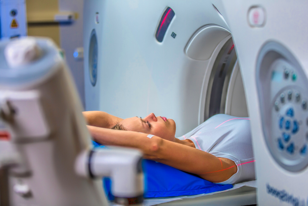
Computed Tomography (CT) scanning technology emits a series of narrow beams through the human body as it moves through an arc and collects data that can then be used for medical and diagnostic purposes.
As a result of the underlying technology, CT scans exhibit many of the same imaging characteristics as traditional x-rays. Images produced by a CT scanning machine can show both soft and hard tissues such as bones, organs, or even the movement of blood within blood vessels. More importantly, this allows doctors to see what is going on inside a patient's body in great detail without having to rely on surgery or other invasive optical instruments.
CT scans can often be used in conjunction with contrast dyes or imaging agents to enhance the quality and fidelity of certain imaging applications such as capturing specific organs or fine details within the cardiovascular system.
CHAPTER 6
How Long Does A CT Scan Take?
How long does a CT Scan take?
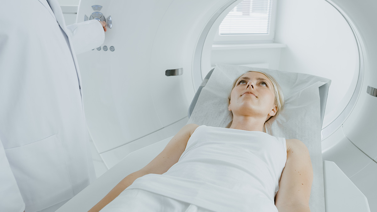
Generally, the average CT scan takes about 5-10 minutes in the scan room when no contrast agents are necessary. When IV contrast is required, you can expect to add an additional 10-15 minutes to the overall procedure. Oral contrast for an abdominal or abdominal and pelvic CT can add an hour or 2.
How Long Does Each Type of CT Scan Procedure Take?
CT scan procedures will vary based on the individual body part or system that requires imaging. Depending on the target, intravenous or oral contrast agents may be necessary. These materials take time to propagate through the body and reach peak accumulation in the targeted area. Depending on the specific body part and method of contrast administration, the timing can vary drastically.
Abdominal or Pelvic CT Scan
2-10 minutes (scanning time); up to 2 hours procedure time if oral contrast used
Chest CT Scan
2 minutes (scanning time); up to 30 minutes when IV access is needed for IV contrast
Low-dose Lung Cancer CT Scan Screening
Spinal CT Scan
2 minutes (scanning time) up to 1 hour procedure time if spinal tap needed to inject contrast into the spinal canal.
The 4 Most Important Factors Affecting CT Scan Times
The precise timing of a CT scan is critical for the success of the procedure. Depending on the body part being scanned, specific preparatory steps may be required that can add to the procedure time. For example, if contrast agents are needed, they must be administered a certain amount of time before the actual scanning procedure to ensure peak visual enhancement.
Certain body parts, organs, and internal anatomical structures require detailed protocols to image. These may require multiple-pass, multiphasic scanning that may add up to 10 minutes to scan time.
Although computer reformatting and enhancement of images does not require more imaging time, it may add time to the window in which the exam is ready for interpretation by a radiologist.
The use of contrast materials will often result in a longer overall imaging procedure times. Depending on the exact CT scanning procedure needed, that increase in time can range from an additional 10 to 15 minutes to as much as 2 hours.
When it comes to contrast dyes, optimal timing is everything. If a CT scan is performed before the contrast materials have had time to accumulate at the target site, the image quality will be affected. Likewise, if the CT scanning procedure is performed after peak dye accumulation, imaging quality will also diminish.
Contrast agent timing will depend on how it is administered and which body parts require imaging.
CT scanning equipment was first introduced in the 1970s. Since that time, scanning equipment has significantly improved, mirroring the overall exponential growth of computing power. Compared to these old machines, many newer CT scanning machines are 7 orders of magnitude faster in regards to imaging speed. What this means is that newer machines can produce high-quality CT scan images in far less time than older machines. The apparent benefit of the rapid increase in the raw imaging speed of CT machines means less time spent on the CT scanning table, and thus, less time spent getting a CT scanning procedure completed.
CHAPTER 7
CAT Scan vs. CT Scan: What's The Difference?
CT vs. CAT Scan: What's the Difference?
Many imaging patients are confused by the terminology used for Computed Tomography (CT) technology in the marketplace today. Imaging facilities often refer to CT scanning technology as “CT scans” or “CAT scans.” “CT scan” is simply an abbreviation of “Computed Tomography scan,” as mentioned above. “CAT,” meanwhile, is an abbreviation for “Computerized Axial Tomography.”
In practice, there is no functional difference between the two terms. They refer to the same procedure and can be used interchangeably. When CT scanners were first introduced into the medical field in the 1970’s they were referred to as EMI machines after the company, EMI Company, that first developed the technology.
So why the difference in terminology?
We’re not quite sure, and we may never know. However, “CT scan” is more commonly used today and has supplanted “CAT scan” in most medical publications.
CHAPTER 8
Are CT Scans Safe?
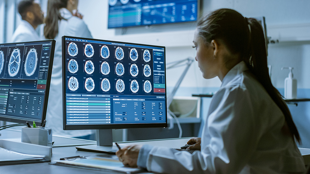
The CT scan is widely used throughout the world as a life-saving diagnostic imaging tool. Every year, nearly 70 million exams are ordered by doctors in the United States alone. Whether for routine screenings or for the rapid imaging and diagnosis of deadly diseases and injuries, CT scans have been credited with saving countless lives.
Nonetheless, many patients still feel concerned about the potential side effects of undergoing a CT scan procedure. More specifically, there is a widespread concern amongst the general public that CT scans may increase their odds of developing cancer.
Despite this concern amongst patients, many doctors believe CT scans to be safe and the potential carcinogenic effects to be negligible. Furthermore, when evaluated from a cost-risk perspective, most medical professionals agree that CT scans are well worth the risk in most cases. With proper risk mitigation, such as the use of protective equipment and adequate calibration, CT scans are much less dangerous than spending time on a tanning bed.
Will CT Scans Increase My Risk of Cancer?
CT scanners are based on x-ray technology, which itself is built around the use of x-ray and gamma radiation to visualize internal body structures such as bones and organs.
X-rays and gamma rays are officially classified as a carcinogen by the World Health Organization’s International Agency for Research on Cancer. What this means is that CT scans are known carcinogens. They do increase a person’s risks of developing cancer. However, this doesn’t necessarily mean that a CT scan is dangerous per se. After all, people get x-rays all the time to diagnose everything from broken bones to dental cavities.
What makes CT scans notable is the relatively higher dosage of radiation each patient receives per session in comparison to a traditional x-ray examination. After all, a CT scan is composed of hundreds of individual x-rays taken at different angles to generate three-dimensional imaging data.
How Safe Are CT Scans Really?
Despite the legitimate concerns that stem from high doses of radiation, many studies have shown CT scans to be relatively safe and the bump in cancer induction to be relatively minor.
Depending on the exact CT scanning procedure, the actual radiation exposure experienced by most patients is usually no more significant than spending a day in the summer sun. An intensive chest CT scan, for example, typically results in between 4 and 7 millisieverts (mSv). A simple, quick CT screening can be as low as 1.5 mSv.
To put the risk posed by an intensive CT scan in context, a single chest CT scan executed with today’s equipment and standards would represent a nominal risk of 0.0025%, or a 1 in 4,000 chance of developing cancer. Because the lifetime risk of dying as a result of cancer is already 1 in 4, or 25%, the added risk of a single, intensive chest scan is statistically negligible.
In and of themselves, CT scans pose only a negligibly higher risk to patients when it comes to developing cancer as a result of exposure to ionizing radiation. However, the real danger to patients is the overprescription and overuse of CT scans. While a single CT scan poses no significant threat, multiple CT scans one after the other can become a significant concern with regards to cancer risks.
The American College of Radiology recommends limiting lifetime diagnostic radiation exposure to 100 mSv to minimize the risks of developing imaging-induced cancer. That’s about 20 to 25 intensive chest CT scans.
Most people will never receive even close to 20 to 25 chest scans until much, much later in life. By the time a person reaches late adulthood, the risks posed by age are far, far more critical than the dangers posed by diagnostic imaging scans. That’s because even if a patient were to develop cancer as a result of receiving too much diagnostic radiation, cancer would still take decades to develop into a life-threatening disease. For patients over the age of 65, the potential preventative benefits of a CT scan often far outweigh any carcinogenic concerns. However, patients under the age of 65 should be careful to maintain a lifetime dosage well below 100 mSv if possible.
How Much Radiation Does A CT Scan Expose You To?
Radiation exposure as a result of a CT scan depends on the type of CT scan being performed and the part of the body that is targeted for imaging purposes. In general, imaging of peripheral parts of the body, such as the legs, feet, arms, and hands results in less radiation exposure while imaging of the body’s core results in more radiation exposure.
To give an idea of how much radiation exposure is associated with standard CT scanning procedures, see below:
- Chest CT Scan: 4-8 mSv
- Head CT Scan: 2 mSv
- Abdominal CT Scan: 8 mSv
- Average US Background
- Radiation: 3.1 mSv
How Can You Minimize Radiation Risk During CT Scans?
One of the best ways to minimize radiation risks is to avoid getting a CT scan if it is not needed. Most doctors will not prescribe a CT scan unless it is necessary. For simple bone imaging, for example, a traditional two-image x-ray may be sufficient, thereby drastically reducing your exposure to ionizing radiation.
Another important way to minimize radiation risks when a CT scan is necessary is to choose an imaging facility with the latest, most modern equipment and certified operators. Modern CT scans are designed to produce higher quality images with less radiation and expose patients to less radiation overall when operated by trained radiologist technicians.
Finally, one of the most obvious ways to avoid excessive CT scanner radiation is to utilize the protective equipment provided by most certified imaging facilities. Depending on the part of the body intended for imaging, imaging clinic often provide lead-lined vests and other pieces of protective gear to reduce a patient's overall radiation exposure.
CHAPTER 9
Are There Open CT Scanners?
How Can I Manage Claustrophobia
and Anxiety During A CT Scan?
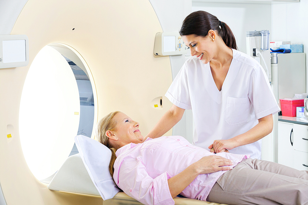
First of all, there are no open CT scanners.
Many patients confuse CT scanners with Magnetic Resonance Imaging (MRI) machines which do offer a so-called “open” version that is suitable for those with claustrophobia, or a fear of enclosed spaces.
What Is Claustrophobia?
Claustrophobia is an anxiety disorder triggered by a variety of environmental stimuli and often results in panic attacks. For those with claustrophobia, small enclosed spaces, or spaces with no windows or doors can cause in sufferers an overwhelming sense of suffocation or fear of restriction. According to Phobias: A Handbook of Theory, Research and Treatment by Graham C. Davey, as much as 5 to 7 percent of the world’s population suffers from some degree of claustrophobia.
Why Is Claustrophobia A Problem During CT Scans?
Claustrophobics often find the prospect of undergoing a CT scan, particularly lengthier CT procedures, a tricky proposition. Many patients with claustrophobia will outright refuse to undergo an important diagnostic imaging scan of any kind, including CT scans and MRIs. As many as 20 percent of patients will skip their imaging procedure as a result of a fear of enclosure and restriction.
Claustrophobia can also cause problems during a CT scan. Nervous, fidgeting patients can make obtaining a high fidelity image difficult.
How To Manage Claustrophobia and Anxiety During a CT Scan?
For patients with mild or manageable claustrophobia as well as general anxiety or antipathy towards medical imaging procedures, there are some easy techniques to help with the process.
For many people, feeling heard goes a long way towards feeling better about a particular medical procedure or treatment. If you have any concerns about a CT scan, talk to your doctor, radiologist, or the radiologist technician. These professionals will be able to talk you through the procedure and provide assurance at the moment.
Some imaging facilities provide patients with the options designed to alter the scanning room environment from soothing music or dim lighting to pictures and paintings of peaceful scenes on the walls.
While usually unnecessary for standard CT scanning procedures, sedatives can sometimes be given to patients who struggle with anxiety or specific medical phobias that could interfere in the scanning process.
Many patients are aware of the best ways to self-prepare for a potentially stressful situation. Whether it’s prior meditation, bringing a friend along, or relaxing in a hot bath beforehand, taking the time to prepare yourself mentally proactively can go a long way towards reducing your overall anxiety during the actual CT scan.


