Arthrograms: A Guide to Diagnostic Joint Imaging
Millions of American both old and young struggle with pain or discomfort in their joints every day. Whether because of a sports-related injury, trauma, or just the slow wearing away of cartilage, joint disorders and joint pain are widespread.
Before a physician can begin prescribing medication, a treatment regimen, or surgical procedure they must first diagnose the cause of the pain and discomfort. This is where arthrograms can help.
What is an Arthrogram?
Despite being something of a tongue-twister, the concept of an arthrogram is quite simple and straightforward. Traditional imaging technologies, such as CT scans, and traditional X-rays, are good for producing interpretable data of bones and MRI exams are good for producing interpretable data on most soft tissues, they may be less effective at imaging soft tissues within the joints. This is a huge obstacle to treating joint pain and joint disorders. You cannot treat what you cannot see. Thankfully, arthrograms make imaging the soft tissue of the joints possible.
An arthrogram is a test that combines traditional imaging techniques with special contrast dyes that make soft tissues such as ligaments, tendons, muscles, cartilage, and joint capsules visible. Essentially, an arthrogram is the combination of an imaging technology, such as an x-ray, with contrast materials such as dyes, water, and air injected directly into the joint rather than into a vein as contrast is usually administered.
Contrast materials are injected into the joints to make them visible to conventional imaging machines. The actual process of injecting dyes and other contrast agents can be precisely guided through two distinct methods: a fluoroscopy or an ultrasound. Fluoroscopy procedures utilize real-time x-rays to help guide the needle trajectory. Ultrasound procedures use images produced by sound waves to plan and guide the needle's path into the joint.
Arthrogram Diagnostic Imaging vs. Traditional Diagnostic Imaging
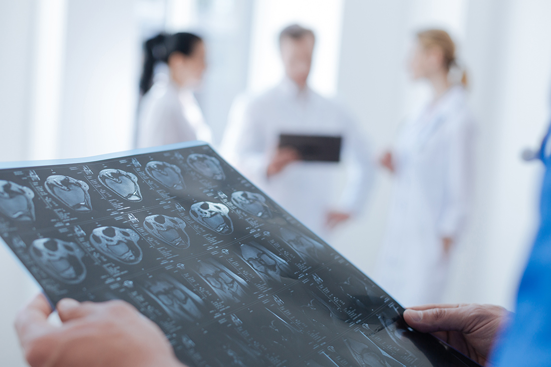
Because arthrograms are a series of images taken of a joint with the aid of contrast dyes, the imaging technique itself can vary. Arthrogram procedures can be executed using almost any traditional imaging technique including magnetic resonance imaging (MRI) scans, CT, and traditional X-rays. While a conventional diagnostic image is typically taken to study the static, hard structures of the body, for example, the bones, arthrograms are designed to study the soft tissues in a joint.
"An arthrogram is a test that combines traditional imaging techniques with special contrast dyes that make soft tissues such as ligaments, tendons, muscles, cartilage, and joint capsules visible."
--- DR. CRISTIN DICKERSON, MD
A Step-By-Step Breakdown of the Arthrogram Procedure
An arthrogram generally takes about 30 minutes to an hour or more depending on the imaging technology utilized and whether multiple diagnostic procedures are required. It is not uncommon for an arthrogram x-ray to be combined with an MRI or CT scan.
Step 1: Initial Consultation
Every arthrogram should begin with an initial consultation to discuss the potential risks and benefits of the procedure. Patients will be asked about any known allergies, especially known allergies to common ingredients in many dyes and contrast agents.
Step 2: Preparation
Once the initial consultation and attendant paperwork are completed, it is time to begin the procedure. The medical professional in charge of the procedure will clean and prepare the skin over the target joint with disinfectants and soap. Clean towels are then draped over the joint followed by an application of local anesthesia to eliminate any pain.
Step 3: Contrast Injection
After the initial preparatory work, the next step is to inject contrast materials into the joint. A needle is inserted into the joint with the guidance of a fluoroscopy or ultrasound equipment followed by the injection of contrast agents.
In some instances, doctors will also remove a sample of joint fluids for testing purposes or to allow the injection of more contrast materials. Once injected, the doctor may instruct you to move your joint to help spread the contrast dyes around more evenly in the joint.
Step 4: Arthrography
The actual process of imaging the joint typically takes place very quickly after the injection of contrast agents before they diffuse into adjacent tissues and structures. Images and data can be extracted using x-rays, MRI, and/or CT. When multiple techniques are used, the radiologist may inject a tiny amount of epinephrine to slow down the diffusion of dyes into unintended areas. When an arthrography is used with an MRI or CT, it can sometimes be referred to as an MRI arthrography or CT arthrography respectively.
Step 5: Recovery
Patient recovery may involve up to 12 hours of rest for the affected joint or joints followed by 1 to 2 days of avoiding strenuous activities. Ice, pain medication, and bandage wraps are rarely necessary after a procedure to cope with swelling and pain.
“Do Arthrograms Hurt?” and Other Common Concerns
Arthrograms are relatively pain-free when local anesthetics are used. There may be a grinding or grating sensation as well as swelling, redness, and tenderness within 24 hours of the procedure. This is normal. Due to the nature of the dyes and the penetration of the needle for the contrast injection, there is a very small risk of an allergic reaction, bleeding in the joint from the injection, or infection.
How Much Do Arthrograms Cost?
Costs will vary widely from one geographic location to another and even from one clinic in a city to another clinic in the same city. In Texas, an arthrogram can cost several thousand dollars. At Green Imaging our fees for an arthrogram are typically around $1000 total.
The Green Imaging Difference
Choosing the right diagnostic exam for you takes a little time, patience, and research. Green Imaging can help. At Green Imaging, we are all about transparency and affordability. With Green Imaging you can save between 50 and 80% of your out-of-pocket costs for MRI, CT, ultrasounds, and other high-quality imaging services. Affordable MRIs start for as low as $250, compared with $1,600 at other imaging facilities in the Houston, TX area.
Don’t pay secret rates for an arthrogram. Go Green Imaging instead!


