Cardiovascular diseases are the first and foremost prominent causes of death in not only the United States but the world at large. In 2015 alone, 17.7 million people died from cardiovascular-related illnesses, complications, and diseases. An additional 70 million Americans suffer from cardiovascular diseases to some extent. Of these deaths caused by cardiovascular diseases, a large percentage are due to coronary heart disease, or coronary artery disease brought about by poor diets, obesity, and lack of adequate physical activity.
Many patients suffering from coronary heart disease are unaware of their conditions and, as a result, end up in the emergency room with little time to spare. In general, health literacy in the United States and around the world is quite poor.
That’s where diagnostic imaging comes into play. Not only does it play a key role in the early diagnosis, treatment, and monitoring of cardiovascular disease, it is increasingly used in emergency rooms to rapidly assess patients experiencing cardiac arrest or distress.

What is an Echocardiogram?
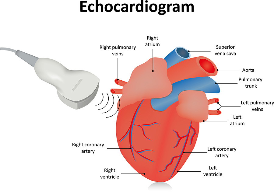
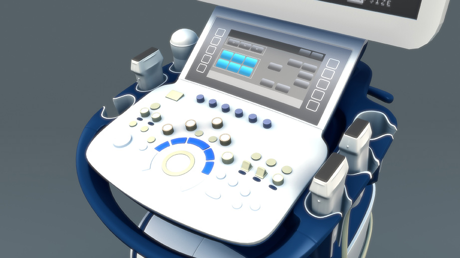
An echocardiogram, or echo test, is an ultrasound-based diagnostic imaging application used to image in 2D and 3D structures and movement of the heart and arteries. Much like a traditional ultrasound used to peer into an expectant mother’s womb, an echocardiogram utilizes sound waves to visualize the heart.
Why Might You Require an Echocardiogram?
Echocardiograms are often standard procedure in many emergency rooms where they are used to assess patients with cardiac distress. They can rapidly produce a real-time visualization of not only the size and shape of the heart and surrounding structures but also movement. This is critical for making a quick, and more importantly, accurate diagnosis that could potentially save a patient’s life in time-sensitive situations.
Outside of the emergency room, echocardiograms are commonly used as a diagnostic tool. After chest x-rays and electrocardiograms, echocardiograms are the third most popular diagnostic procedure for patients with suspected heart conditions. This is because echocardiograms provide a wealth of information quickly, noninvasively, and with minimal discomfort for the patient.
Information provided by an echocardiogram includes the size and shape of a patient’s heart, size of internal chambers, pumping capacity, and the exact location and extent of any damage to heart tissues. When combined with pulsed or continuous-wave doppler technologies, echocardiograms can also provide important data about blood flow throughout the body. Echo procedures can help a doctor diagnose high blood pressure, a leaky heart valve, congenital heart defects, or even impending heart failure.
The key to successfully treating heart disease is to catch the problem early. Early diagnosis and treatment are critical. Patients who suspect that they may have a cardiovascular condition should have an echocardiogram done as soon as possible.
5 Signs You Should Get an Echocardiogram Right Now
1. Chest Pain
Chest pain, or angina, is a red flag that something isn’t right with the heart. While it may just be acid reflux from the stomach into the esophagus, persistent episodes of angina that last 5 to 10 minutes before subsiding could indicate heart disease and an impending heart attack. When in doubt, get it checked out.
2. Pain In Arms, Neck, Back, or Jaw
By itself, pain in the arms, neck, back, or jaw can be due to a variety of other factors, including pulled or strained muscles, pinched nerves, or temporomandibular joint (TMJ) disorders. However, when pain in the arms, neck, back, or jaw is accompanied by other traditional signs of cardiac distress, such as sweating, nausea, fatigue, and chest pain, it’s time to get your heart checked immediately.
3. Shortness of Breath, Fatigue, Weakness
Unlike men who tend to experience angina or heart pain, during cardiac distress, women often experience other symptoms along with chest discomfort, such as shortness of breath and fatigue. Additionally, weakness, particularly in the limbs, is a sign that blood isn’t getting through as a result of narrowed or restricted blood vessels.
4. Irregular Heart Beat
An irregular heartbeat or changes in your heart rhythm could be indicative of endocardium, or a bacterial infection of the heart, dilated cardiomyopathy, or weak heart muscles, valvular heart disease, or other forms of heart arrhythmias. Each of these is bad news and can be diagnosed with an echocardiogram.
5. Swollen Arms, Legs, Feet, or Ankles
Edema, or swelling caused by excessive fluids trapped in a patient’s tissues, is indicative of underlying heart disease.

Types of Echocardiograms
Several types of echocardiograms exist. All use ultrasound sonar technology, in which sound waves passed through the chest are used to image a patient’s heart and arteries. However, different types of echocardiograms have different applications and uses.
Transthoracic Echocardiogram
This is the most common kind of echocardiogram. Typically, an ultrasound wand, or transducer, is placed on a patient’s chest. The device transmits sound waves through the chest cavity to create images and data of the heart and heart walls.
Transesophageal Echocardiogram
Sometimes, a standard transthoracic echocardiogram cannot create images accurate enough for a doctor to use. In this case, a transducer attached to a thin tube may need to be guided into a patient’s esophagus to get as close to the heart as possible. This allows for higher fidelity, more accurate images.
Stress Echocardiogram
Stress echocardiograms are given in conjunction with a stress test to assess the heart when it is working hard and beating quickly. Stress echocardiograms often involve walking or running on a treadmill or pedaling on a stationary exercise bike. It is most often used to diagnose coronary heart disease.
3D Echocardiogram
New advancements in transthoracic imaging technologies allow doctors to generate three-dimensional data and pictures of a patient’s heart. This is typically used to help doctors and surgeons plan heart valve surgery and other incredibly complex operations in which precise information is key.
Fetal Echocardiography
A fetal echocardiogram is a test aimed at assessing an unborn child’s heart. Like a traditional ultrasound, a fetal echocardiogram involves moving the transducer over a pregnant women’s belly. This test is typically done between 18 and 22 weeks of pregnancy to check for congenital heart disorders or other potential cardiovascular problems.


